Hair Cloning Research - Bernstein Medical - Center for Hair Restoration
About Header Image

Scientific research is often the quintessential example of taking something apart to learn how it works. A team of researchers has used that age-old technique to unwind the complex process by which embryonic cells organize into functional skin that includes “organoids” like hair follicles. By untangling this biological mystery, they were able to develop a model that could potentially lead to hair regeneration treatments and other advances in regenerative medicine. The study — “Self-organization process in newborn skin organoid formation inspires strategy to restore hair regeneration of adult cells” — was published in the August 22nd, 2017 issue of the journal PNAS. ((Lei M, Schumacher LJ, et al. Self-organization process in newborn skin organoid formation inspires strategy to restore hair regeneration of adult cells. Proceedings of the National Academy of Sciences. 114. 201700475. 10.1073/pnas.1700475114.))

Background

Scientists have long known that embryonic cells organize and form bodily organs like the heart, liver, and skin, but understanding the details behind this spontaneous process of “self-organization” was a challenge. It has been understood that the DNA code produces chemicals that enable “cross-talk” between cells as they go through several stages in forming 3-dimensional, functional organs. But what these stages represented, and which specific molecules were involved in this communication, needed to be understood.

The Study

The researchers set out with two goals in mind: 1) to describe the conditions that enable a group of cells to self-organize into skin organs and 2) to describe transplant techniques that allow these skin organs to grow normal, functional hairs.

To achieve their first goal, the researchers started with populations of individual “dissociated” epidermal and dermal cells from newborn mice. They then combined these cells in a 3-D cell drop and observed the cells’ interactions. At every stage, they measured how the cells behaved and which proteins and molecules were present to promote or inhibit certain processes. The dissociated cells self-organized into functional skin through a complex 5-stage process:

  • Stage 1: Cells form aggregations
  • Stage 2: Aggregates form cysts
  • Stage 3: Cysts fuse to form epidermal “planes”
  • Stage 4: Small epidermal planes merge to form a larger, multi-layered plane of embryonic skin
  • Stage 5: Embryonic skin forms “placode” structures that can develop into hair follicles

When the Stage 5 cultured skin was transplanted to the back of a hairless mouse, it grew robust hair follicles.

Self-organization process in newborn skin organoid formation inspires strategy to restore hair regeneration of adult cells
(A) The experiment design. (B) Images showing the self-organization process. (C) Diagram of the stages of new skin formation. (D) Robust hair regeneration seen with cells from newborn mice but not adults. (E) Adult cells form only small aggregates. (F) Aggregate size with cells from newborn mice vs. adults. (G) Schematic showing how self-organization in adult cells stops before growth is complete. Image c/o PNAS

With a better understanding of these processes using embryonic cells, they went about attempting to induce this same process in adult mouse skin cells. Adult cells on their own formed only a few small aggregates which did not grow. To confirm the significance of newborn dermal cells and the chemicals that cause them to self-organize, the researchers experimented by first combining newborn dermal cells and adult epidermal cells and then combining newborn epidermal cells and adult dermal cells. The population that contained newborn dermal cells formed numerous hairs, while the population with newborn epidermal cells formed very few hairs.

Findings

  • Researchers identified several different classes of molecules that are required to induce the transitions between the various stages of skin development
  • The transition periods were not discrete events, but instead occurred over time and were dependent on the presence of the “communication” molecules
  • Inhibiting or promoting the key communication molecules can suppress or accelerate the phase transition process
  • Adding the communication molecules to adult cell cultures at the appropriate times can induce the adult cells to form functional skin complete with a robust number of hair follicles
  • It is both the progression of the phases and the presence of the molecular signals that, together, form the key to self-organization

Conclusion

The researchers behind this study sought to achieve two complicated tasks: to explain how cells self-organize and to induce self-organization in cells which had lost that capability. Only through painstaking experimentation were they able to untangle some of the mystery behind how these embryonic cells transform from a group of individual cells into fully-formed skin complete with hair follicles. This effort paid off, as they were able to apply the newfound knowledge to populations of adult cells. The capability to induce robust hair follicle growth in adult skin is a significant achievement.

For now, we must be content that the adult skin that was cultured in the lab needed to be grafted onto a healthy host. Next steps would be to determine how to use the same or similar process to induce hair follicle growth directly in the skin. Much more research must be done in that regard. However, the implications of this technique for the field of regenerative medicine may be substantial, as scientists explore the ability to regenerate not only skin organs but other body organs in the future.

Posted by

New research published in the journal PLoS One found that embryonic stem cells can be used to form a type of cell that induces new hair follicle growth, and that these cells promote robust hair growth when implanted into mice.

Background

Dermal Papilla (DP) cells play a role in new hair follicle formation and in the growth of new hair. Because of this role, it was hoped that DP cells grown in the laboratory (i.e., grown in culture) could form the basis of a treatment for genetic hair loss. However, it turned out that these cultured DP cells lost their hair follicle-inducing potential too quickly to be useful in treating hair loss.

New Research

Now, however, new research has found that human embryonic stem cells (hESCs) can generate cells that are functionally equivalent to DP cells. ((Gnedeva K, Vorotelyak E, Cimadamore F, Cattarossi G, Giusto E, Terskikh V.V, Terskikh A.V. Derivation of hair-inducing cell from human pluripotent stem cells. PLoS One. 2015 Jan 21;10(1)) Like DP cells, these functionally equivalent cells can induce hair follicle formation just as readily as DP cells. But more significantly, unlike cultured DP cells, they do not lose their potential to induce hair follicle formation when grown in the laboratory. This discovery represents an important advance in developing a hair cloning technique to cure pattern baldness.

Posted by

New research published in the journal Developmental Cell has confirmed the importance of dermal sheath stem cells in maintaining the hair growth cycle. ((Rahmani W, et al. Hair Follicle Dermal Stem Cells Regenerate the Dermal Sheath, Repopulate the Dermal Papilla, and Modulate Hair Type. Dev Cell. 2014 Dec 8;31(5):543-58.)) These cells, located around the lower portion of growing follicles, form the basis of an experimental treatment, being developed by Replicel Life Sciences, Inc., to regenerate hair-producing follicles. If successful, the treatment will be a game-changer for the hair restoration industry.

Colony of self-renewing dermal sheath cellsColony of self-renewing dermal sheath cells

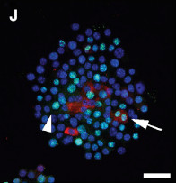
The study, published in December 2014, sought to confirm what had been indirect evidence of a type of stem cell residing in the dermal sheath (DS) that was said to replenish dermal papilla (DP) cells. The authors of the study suggest that they now have definitive evidence that new DP cells are derived from stem cells in the dermal sheath “cup” (DSC). This development clarifies the relationship between the DS and the DP and confirms that DSC cells play a critical role in hair follicle regeneration by repopulating the dermal papilla cells at the end of the telogen (resting) phase of the normal hair cycle.

Importance of the Dermal Sheath Cup Cells

The number of dermal papilla (DP) cells in a hair follicle has been found to be a determining factor as to when the anagen (growth) phase of the hair cycle is initiated. ((Chi W, Wu E, Morgan BA, et al. (2013). Dermal papilla cell number specifies hair size, shape and cycling and its reduction causes follicular decline. Development 140, 1676–1683.)) The gradual loss of DP cells over time results in a longer delay in the onset of the anagen phase; a longer telogen (resting) phase; and a hair follicle that shrivels and eventually disappears. This process, called miniaturization, plays out over multiple hair cycles and has been shown to be the primary contributor to androgenetic alopecia and eventual baldness. ((Randall VA. (2008). Androgens and hair growth. Dermatol. Ther. 21, 314–328.))

While dermal sheath cup (DSC) stem cells are known to be long-lived and self-renewing, it is not fully understood how they replicate or why the pool of DSC cells becomes depleted over time. We do know, however, that the gradual loss of DSC cells results in a failure to produce the necessary number of DP cells. And without enough DP cells to trigger the anagen phase, the follicle begins to miniaturize. It is clear that maintaining the population of DSC cells after each iteration of the hair cycle is very important in preserving and maintaining healthy and mature terminal hairs.

Replicel Reacts to the Study

The new data confirming the importance of the dermal sheath cup (DSC) cells was celebrated by researchers and executives at Replicel Life Sciences, Inc., who have been studying this issue for over a decade. Replicel is set to start phase II clinical trials of RCH-01, their proprietary treatment for androgenetic alopecia.

In the RCH-01 treatment, cloned DSC cells are injected into balding areas of the scalp where they are expected to reverse miniaturization and regenerate healthy, hair-producing follicles. Phase I trials of RCH-01, the results of which were published in 2012, showed that the treatment could produce promising results and that it was safe to administer. Six months after patients were treated with RCH-01, overall hair density increased by an average of 11.8% in ten patients out of 16. In two patients, overall hair density increased by more than 19%. There were no significant adverse safety events recorded. ((Lortkipanidze, N. Safety and Efficacy Study of Human Autologous Hair Follicle Cells to Treat Androgenetic Alopecia. In Clinicaltrials.gov. Retrieved July 26, 2012.)) Phase II clinical trials are set to begin in 2015, with data collection continuing for 39 months.

Through a 2013 agreement with Replicel, Japanese cosmetics giant Shiseido may introduce RCH-01 into the Asian market as early as 2018.

Image c/o Developmental Cell 31, 543–558, December 8, 2014 ª2014 Elsevier Inc.

Posted by

Dr. Claire Higgins and her colleague Dr. Colin Jahoda have published an overview of hair cloning and the challenges scientists face in attempting to develop hair regeneration therapies for androgenetic alopecia or common balding. The article, published in Hair Transplant Forum International, points to two central problems in developing a hair loss therapy. The first is the difficulty in getting dermal papilla cells in humans to self-aggregate and form hair follicles and the second is the inability, thus far, of scientists to generate normal hairs and follicles.

Higgins and Jahoda describe how it has been known for decades, through the work of Lille and Wang and others, that rat dermal papillae self-organize into new hair-producing follicles when they are injected or grafted into the skin. Human dermal papilla cells, on the other hand, have never exhibited what they call the “aggregation phenomena,” and instead they disperse in the skin in what appears to be a wound healing mechanism. In fact, human papillae, when grown in a laboratory culture, can act as “mesenchymal stem cells” and differentiate into a variety of cell types.

While multiple efforts to induce dermal papillae to form new hair follicles have failed, the research that Higgins and Jahoda have published on hair follicle neogenesis has resulted in a new technique to do just that. The success of the 3-D culturing of dermal papillae to induce hair follicle neogenesis was a breakthrough in that the scientists have found a way to improve the intercellular communication that is essential to inducing follicle growth.

Having made significant progress in improving this vital communication link between dermal papillae cells, scientists still have to contend with a series of obstacles that stand in the way of a hair cloning therapy for human hair loss. One such problem is the quality of hairs that they have been able to grow using the hair follicle neogenesis technique. The hairs they have successfully produced have been small and have grown in non-uniform direction. Another unanswered issue is how long the hair follicles will grow and whether or not they exhibit the cyclical hair follicle growth patterns of a typical human hair follicle. The ability to reproduce significant quantities of normal hair will continue to be the central focus of research going forward.

Bookmark our Hair Cloning Research page to stay on top of developments in this field

Posted by

Progress towards hair cloning may have just have shifted up another gear thanks to scientists at the University of Pennsylvania and the New Jersey Institute of Technology. The breakthrough study published January 28th, 2014 is the first to show the successful transformation of adult human skin cells into quantities of epithelial stem cells necessary for hair regeneration.

The researchers, led by Dr. Xiaowei “George” Xu, started with human skin cells called dermal fibroblasts, then transformed those into a type of stem cell called induced pluripotent stem cells (iPSCs). These were then transformed into epithelial stem cells (EpSCs). This important step had never been achieved before in either humans or mice. The epithelial stem cells were combined with mouse dermal cells, that can be induced to form hair follicles, and then grafted on a mouse host. The epithelial cells and dermal cells then grew to form a functional human epidermis and follicles structurally similar to human hair follicles. The exhibits that accompany the study include photographic evidence of human hairs.

Figure 5 - Generation of folliculogenic human epithelial stem cells from induced pluripotent stem cellsHair shafts (arrowheads) formed by induced pluripotent stem cell-derived epithelial stem cells compared to mouse hair (arrows). — Credit: Ruifeng Yang, Perelman School of Medicine, University of Pennsylvania

The main breakthrough in the study came when the research team carefully timed the addition of growth factors to the iPSCs. Previous research showed the ability for iPSCs to be transformed into a common type of cell found in the skin called keratinocytes. By timing the addition of the growth factors, they were able to turn over 25% of the iPSCs into epithelial stem cells in a little more than two weeks. This “mass production” of epithelial stem cells holds tremendous promise for the development of a hair regeneration treatment. On this development, Dr. Xu said, “This is the first time anyone has made scalable amounts of epithelial stem cells that are capable of generating the epithelial component of hair follicles.”

As noted in a University of Pennsylvania press release on the news, there are two types of stem cell that are critical in hair follicles: epithelial stem cells and dermal papillae. While this study only achieved success in the creation of epithelial stem cells, we have extensively covered Dr. Angela Christiano’s ground-breaking research into the induction of dermal papillae into hair follicles (a process she calls hair follicle neogenesis).

“When a person loses hair, they lose both types of cells. We have solved one major problem, the epithelial component of the hair follicle. We need to figure out a way to also make new dermal papillae cells, and no one has figured that part out yet,” said Dr. Xu.

Once that it is done, we must also find a way to have the epithelial and dermal components of the follicle interact before one will be able to produce cosmetically useful hair. But with each successive breakthrough, the time when a scientist can use hair cloning techniques to regenerate human hair, and the surgeon can implant them into a person’s scalp, draws ever closer.

Reference
Yang R, Zheng Y, Xu X. Generation of folliculogenic human epithelial stem cells from induced pluripotent stem cells. Nature Communications. 2014.

Posted by

Q: I read, with considerable interest, your excellent article on the latest in Dr. Angela Christiano’s work on follicular neogenesis. It seems to me that the next questions we should be asking are: when will testing begin on human subjects and when might her research develop into a hair cloning treatment that is available to the general public?

A: It is very difficult to determine when this phase of the research might begin and it is even harder to predict when treatment might become available. First, the technology is not quite there. Dr. Christiano showed in her recent paper that changing the environment of skin (fibroblast) cells so that they could form into 3-D cultures enabled them to induce human hair-follicle growth. Although this was a major step towards cloning hair, additional work needs to be done before we will be able to mass produce fully-functioning human hair follicles to the extent needed for hair transplantation.

In addition, research on human subjects requires that experiments meet rigorous federal regulatory standards and these take time to be approved and carried out. Supposing that further study of follicle neogenesis results in a breakthrough treatment for hair loss, this treatment would still require meeting substantial efficacy and safety requirements of the FDA before it would be made available to the public. We will be communicating important developments as they occur through our Hair Cloning Research section and through periodic updates in the Bernstein Medical Newsletter.

Posted by

Scientists from Durham University in the UK have shown for the first time that a lab technique, called a three-dimensional cell culture, can produce spherical structures that are similar to naturally occurring structures in hair follicle formation (called dermal papilla or DP). This breakthrough study by Claire Higgins and Colin Jahoda, published in the June 2010 issue of the journal Experimental Dermatology, ((Higgins C, Jahoda C, et al. Modelling the hair follicle dermal papilla using spheroid cell cultures. Experimental Dermatology 2010; 19: 546–548.)) has the potential to unlock the ability of researchers to develop functional DP cells which can be used in hair restoration techniques such as hair cloning or hair multiplication.

Background

Hair cloning techniques have been theorized for decades. The basic idea is:

  1. a physician takes a sample of skin cells from a patient
  2. dermal papilla cells are extracted
  3. the DP cells are cloned (multiplied) in a laboratory culture (i.e., a petri dish)
  4. the cell formation is then injected back into the patient’s balding scalp where it produces permanent hair that continues to grow

The first three steps are a piece of cake. But that is when the strategy breaks down. When DP cells are grown in a petri dish they exhibit some of the qualities of DP cells in the human body but not all, so injecting this aggregate into the skin fails to lead to hair follicle growth. Something was missing.

In 1991, Wobus, et al published a study in the journal Differentiation ((Wobus AM, Wallukat G, Hescheler J. Differentiation 1991: 48: 173–182.)) that described a new technique for researching cells that in nature exist as clumps or masses of cells. The idea was to suspend a group of cells under a flat surface so that gravity would pull the cells into a droplet. This “hanging drop” method yielded a three-dimensional culture that enabled the study of embryonic stem cells as well as the proteins they produce that allow for intercellular communication.

Having hit the wall with two-dimensional DP cultures, Higgins and Jahoda set out to try Wobus’ concept of using 3-D cultures to study DP cells.

The Study

Higgins and Jahoda harvested eight cell strains of human DP cells taken from scalp hair follicles. These eight strains were cultured in either 35-mm dishes or hanging drop cultures consisting of 3,000 cells each. The cultures were maintained between 30 and 72 hours, then collected and analyzed using immunofluorescence or transcriptional techniques.

Results

The DP cells grown in hanging drop, 3-D cultures exhibited behavior significantly akin to DP in human hair follicles. The 2-D cultures grown in the 35-mm dishes did not.

Conclusion

Without the ability to form functional dermal papilla aggregations, hair cloning was essentially at a dead end. In the 3-D configuration, the aggregated cells were able to communicate with one another and to continue to differentiate as hair follicles. By using Wobus’ 3-D hanging drop technique, Higgins and Jahoda may have unlocked the secret to forming these powerful, but elusive, structures that are critical to the hair growth cycle.

Following this study, more research needs to be performed to induce the spherical cells to initiate the growth of new hair follicles and to develop ways to ensure that the induced hair follicles are immune from the factors that cause genetic hair loss. Should those two riddles be solved, hair loss will have been effectively cured.

Posted by


Browse Hair Restoration Answers by topic:


212-826-2400
Scroll to Top