Dermal Papilla - Bernstein Medical - Center for Hair Restoration
About Header Image

Two new studies researching a class of drugs called JAK inhibitors have shown that oral treatment results in significant hair regrowth in patients with alopecia areata, an autoimmune condition that causes non-scarring patches of localized hair loss. Currently there is no cure for alopecia areata, so the possibility of a safe, effective medication is welcome news for thousands of affected patients.

Background

Last year we wrote about how the two new FDA-approved drugs tofacitinib and ruxolitinib act as inhibitors of the family of enzymes called Janus kinase (JAK). ((Harel S, Higgins CA, Cerise JE, Dai Z, Chen JC, Clynes R, Christiano AM. Pharmacologic inhibition of JAK-STAT signaling promotes hair growth. Sci Adv. 2015 Oct; 1(9): e1500973.)) By inhibiting the JAK enzymes, the drugs disrupt intracellular communication to white blood cells, called “T lymphocytes,” and are thus useful in treating alopecia areata. The JAK inhibitors prevented the onset of the disease and reversed the condition, enabling hair to regrow in areas previously devoid of hair.

The 2015 study we referenced – led by renowned alopecia areata researcher Dr. Angela Christiano – showed that topical application of tofacitinib and ruxolitinib in mice resulted in the rapid transition of hair follicles from the telogen (resting) phase of the hair cycle to the anagen (growth) phase. The same study found that tofacitinib encouraged hair follicle development in clumped human dermal papilla (DP) cells, stem cells that are critical in the development of hair follicles. [1]

The Studies

The two new studies were published in September 2016 in the journal JCI Insight, a peer-reviewed journal dedicated to biomedical research.

Tofacitinib

The study of oral tofacitinib – by Crispin, Ko, et al – was a 2-center, open-label, single-arm trial; the first to systematically examine the efficacy of JAK inhibitors as a treatment for alopecia areata. ((Crispin MK, Ko J, Craiglow BG, Li S, Shankar G, Urban JR, Chen JC, Cerise JE, Jabbari A, Winge MG, Marinkovich MP, Christiano AM, Oro AE, King BA. Safety and efficacy of the JAK inhibitor tofacitinib citrate in patients with alopecia areata. JCI Insight. 2016;1(15):e89776. doi:10.1172/jci.insight.89776.)) In addition to studying alopecia areata (AA) patients with greater than 50% scalp hair loss, they tested the drug on patients with alopecia totalis (AT), which is the complete loss of scalp hair; alopecia universalis (AU), the loss of scalp and body hair; and ophiasis pattern alopecia areata, hair loss localized to the temporal and occipital scalp. After three months on 5mg tofacitinib citrate, 32% showed up to 50% improvement, and 32% showed greater than 50% improvement. When broken down by subtype of the condition, those with AA improved by 70% on average, those with ophiasis improved by 68%, AT by 11.8%, and AU by 10.5%. They found that following cessation of the treatment, all patients experienced a recurrence of hair loss after an average of 8.5 weeks. Additional trials are necessary to determine the optimal dosage regimen for providing the most long-lasting response.

Ruxolitinib

The study of ruxolitinib – by Mackay-Wiggan, Jabbari, et al – was an open-label clinical trial of 12 patients with moderate to severe alopecia areata. ((Mackay-Wiggan J, Jabbari A, Nguyen N, Cerise J, Clark C, Ulerio G, Furniss M, Vaughan R, Christiano AM, Clynes R. Oral ruxolitinib induces hair regrowth in patients with moderate-to-severe alopecia areata. JCI Insight. 2016;1(15):e89790. doi:10.1172/jci.insight.89790.)) The pilot study tested the use of 20mg oral ruxolitinib twice a day for three to six months; this was followed by three months of monitoring the patients without treatment. Despite the small sample size, the results were striking in that 75% of patients showed a strong response to the medication, with hair regrowth over 50%. After treatment, those who responded to the treatment exhibited a 92% reduction in hair loss. Seven of the nine responders achieved greater than 95% hair regrowth. After stopping treatment hair loss resumed; however, it did not reach the level of hair loss that was present before treatment. This proof-of-concept pilot study showed that ruxolitinib is a safe and effective in reversing the balding effects of alopecia areata.

Conclusion

After showing promise in previous research, scientists have now shown that JAK inhibitors have strong potential to cause substantial hair regrowth in people with alopecia areata; a condition that causes hair loss that can be socially awkward at best and cosmetically disfiguring in severe cases. More studies need to go forward in order to determine which of the two drugs – tofacitinib or ruxolitinib – will be the most effective treatment, and what the proper dosage is for long-term treatment. However, we are hopeful that a medication will be developed for broad use in treating alopecia areata patients.

The other major point of interest following the publication of the series of studies is the potential for JAK inhibitors to treat androgenetic alopecia, or common genetic hair loss. One area that is being discussed is the potential for JAK inhibitors, perhaps in the form of a topical treatment, to stimulate the transition of hair follicles from the resting phase to the growth phase of the hair cycle. Christiano’s research is examining the effects of JAK inhibitors on cultured dermal papilla (DP) spheres. If JAK inhibitors can be used to stimulate DP spheres to grow into mature hair follicles, it may enable hair multiplication techniques to become a viable treatment for common baldness.

Posted by

New research published in the journal PLoS One found that embryonic stem cells can be used to form a type of cell that induces new hair follicle growth, and that these cells promote robust hair growth when implanted into mice.

Background

Dermal Papilla (DP) cells play a role in new hair follicle formation and in the growth of new hair. Because of this role, it was hoped that DP cells grown in the laboratory (i.e., grown in culture) could form the basis of a treatment for genetic hair loss. However, it turned out that these cultured DP cells lost their hair follicle-inducing potential too quickly to be useful in treating hair loss.

New Research

Now, however, new research has found that human embryonic stem cells (hESCs) can generate cells that are functionally equivalent to DP cells. ((Gnedeva K, Vorotelyak E, Cimadamore F, Cattarossi G, Giusto E, Terskikh V.V, Terskikh A.V. Derivation of hair-inducing cell from human pluripotent stem cells. PLoS One. 2015 Jan 21;10(1)) Like DP cells, these functionally equivalent cells can induce hair follicle formation just as readily as DP cells. But more significantly, unlike cultured DP cells, they do not lose their potential to induce hair follicle formation when grown in the laboratory. This discovery represents an important advance in developing a hair cloning technique to cure pattern baldness.

Posted by

New research published in the journal Developmental Cell has confirmed the importance of dermal sheath stem cells in maintaining the hair growth cycle. ((Rahmani W, et al. Hair Follicle Dermal Stem Cells Regenerate the Dermal Sheath, Repopulate the Dermal Papilla, and Modulate Hair Type. Dev Cell. 2014 Dec 8;31(5):543-58.)) These cells, located around the lower portion of growing follicles, form the basis of an experimental treatment, being developed by Replicel Life Sciences, Inc., to regenerate hair-producing follicles. If successful, the treatment will be a game-changer for the hair restoration industry.
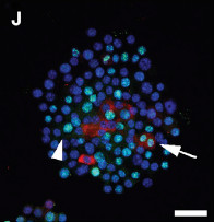
Colony of self-renewing dermal sheath cellsColony of self-renewing dermal sheath cells

The study, published in December 2014, sought to confirm what had been indirect evidence of a type of stem cell residing in the dermal sheath (DS) that was said to replenish dermal papilla (DP) cells. The authors of the study suggest that they now have definitive evidence that new DP cells are derived from stem cells in the dermal sheath “cup” (DSC). This development clarifies the relationship between the DS and the DP and confirms that DSC cells play a critical role in hair follicle regeneration by repopulating the dermal papilla cells at the end of the telogen (resting) phase of the normal hair cycle.

Importance of the Dermal Sheath Cup Cells

The number of dermal papilla (DP) cells in a hair follicle has been found to be a determining factor as to when the anagen (growth) phase of the hair cycle is initiated. ((Chi W, Wu E, Morgan BA, et al. (2013). Dermal papilla cell number specifies hair size, shape and cycling and its reduction causes follicular decline. Development 140, 1676–1683.)) The gradual loss of DP cells over time results in a longer delay in the onset of the anagen phase; a longer telogen (resting) phase; and a hair follicle that shrivels and eventually disappears. This process, called miniaturization, plays out over multiple hair cycles and has been shown to be the primary contributor to androgenetic alopecia and eventual baldness. ((Randall VA. (2008). Androgens and hair growth. Dermatol. Ther. 21, 314–328.))

While dermal sheath cup (DSC) stem cells are known to be long-lived and self-renewing, it is not fully understood how they replicate or why the pool of DSC cells becomes depleted over time. We do know, however, that the gradual loss of DSC cells results in a failure to produce the necessary number of DP cells. And without enough DP cells to trigger the anagen phase, the follicle begins to miniaturize. It is clear that maintaining the population of DSC cells after each iteration of the hair cycle is very important in preserving and maintaining healthy and mature terminal hairs.

Replicel Reacts to the Study

The new data confirming the importance of the dermal sheath cup (DSC) cells was celebrated by researchers and executives at Replicel Life Sciences, Inc., who have been studying this issue for over a decade. Replicel is set to start phase II clinical trials of RCH-01, their proprietary treatment for androgenetic alopecia.

In the RCH-01 treatment, cloned DSC cells are injected into balding areas of the scalp where they are expected to reverse miniaturization and regenerate healthy, hair-producing follicles. Phase I trials of RCH-01, the results of which were published in 2012, showed that the treatment could produce promising results and that it was safe to administer. Six months after patients were treated with RCH-01, overall hair density increased by an average of 11.8% in ten patients out of 16. In two patients, overall hair density increased by more than 19%. There were no significant adverse safety events recorded. ((Lortkipanidze, N. Safety and Efficacy Study of Human Autologous Hair Follicle Cells to Treat Androgenetic Alopecia. In Clinicaltrials.gov. Retrieved July 26, 2012.)) Phase II clinical trials are set to begin in 2015, with data collection continuing for 39 months.

Through a 2013 agreement with Replicel, Japanese cosmetics giant Shiseido may introduce RCH-01 into the Asian market as early as 2018.

Image c/o Developmental Cell 31, 543–558, December 8, 2014 ª2014 Elsevier Inc.

Posted by

Could a hormone that plays a critical role in red blood cell production also play a critical role in hair follicle production? According to a 2010 research report published in the Journal of Dermatological Science, this may be the case.

Erythropoietin Implicated In Hair Growth Regulation

The hormone in question is called Erythropoietin (EPO). It is produced in the kidneys in order to regulate red blood cell production. Recent studies have shown that EPO is also produced in a structure that surrounds and protects a hair follicle, the outer root sheath (ORS). Moreover, other studies have found that the EOP secreted by the ORS seems to target dermal papilla (DP) cells. DP cells play a critical role in regulating hair growth.

Because of these results, researchers have speculated that EPO may affect hair growth by acting on DP cells, but no direct evidence for this had ever been found – until now.

Evidence That EPO Affects Hair Growth in Vitro (Cell Cultures)

Strong evidence of EPO’s direct involvement in hair growth would be the discovery of EPO receptor sites (EPOR) on DP cells and a clear mechanism of how EPO affects changes in a DP cell (called cell signaling); this is exactly what researchers in the Republic of Korea ((Kang BM, Shin SH, Kwack MH, Shin H, Oh JW, Kim J, Moon C, Moon C, Kim JC, Kim MK, Sung YK. Erythropoietin promotes hair shaft growth in cultured human hair follicles and modulates hair growth in mice. J Dermatol Sci. 2010 Aug;59(2):86-90. doi: 10.1016/j.jdermsci.2010.04.015. Epub 2010 May 19.)) have found. Not only did they find direct evidence of EPO receptive sites but they also discovered the critical cell signaling mechanism: phosphorylated EPOR signaling pathway mediators.

In addition to discovering the signaling mechanism, they also showed using cell cultures that EPO causes both dermal papilla to proliferate and hair shafts of human hair follicles to elongate.

While the effects of EPO on DP and hair follicles were compelling, they only occurred in vitro (in cell cultures outside the body) and it is known that cells cultured on a flat surface behave significantly differently than cells that exist in situ, inside the organism (see Higgins and Christiano, Regenerative Medicine And Hair Loss: How Hair Follicle Culture Has Advanced Our Understanding Of Treatment Options For Androgenetic Alopecia).

Evidence That EPO Affects Hair Growth In Situ (In The Body)

In order to better answer the questions of whether and how EPO might directly affect hair growth in situ, the Korean researchers implanted EPO treated DP cells into mice and found that these treated cells not only moved hair follicles from their resting (telogen) phase into an active hair growth (anagen) phase but also prolonged a follicle’s active growth phase.

This is a significant finding since one of the mechanisms of male pattern baldness is DHT susceptible hair follicles entering into progressively longer periods of a telogen (resting) phase relative to an anagen (hair growth) phase. EPO, having the opposite effect on hair follicles, opens the door to treating this type of hair loss with existing EPO analogs and/or developing new erythropoietin biopharmaceuticals.

Posted by

Dr. Claire Higgins and her colleague Dr. Colin Jahoda have published an overview of hair cloning and the challenges scientists face in attempting to develop hair regeneration therapies for androgenetic alopecia or common balding. The article, published in Hair Transplant Forum International, points to two central problems in developing a hair loss therapy. The first is the difficulty in getting dermal papilla cells in humans to self-aggregate and form hair follicles and the second is the inability, thus far, of scientists to generate normal hairs and follicles.

Higgins and Jahoda describe how it has been known for decades, through the work of Lille and Wang and others, that rat dermal papillae self-organize into new hair-producing follicles when they are injected or grafted into the skin. Human dermal papilla cells, on the other hand, have never exhibited what they call the “aggregation phenomena,” and instead they disperse in the skin in what appears to be a wound healing mechanism. In fact, human papillae, when grown in a laboratory culture, can act as “mesenchymal stem cells” and differentiate into a variety of cell types.

While multiple efforts to induce dermal papillae to form new hair follicles have failed, the research that Higgins and Jahoda have published on hair follicle neogenesis has resulted in a new technique to do just that. The success of the 3-D culturing of dermal papillae to induce hair follicle neogenesis was a breakthrough in that the scientists have found a way to improve the intercellular communication that is essential to inducing follicle growth.

Having made significant progress in improving this vital communication link between dermal papillae cells, scientists still have to contend with a series of obstacles that stand in the way of a hair cloning therapy for human hair loss. One such problem is the quality of hairs that they have been able to grow using the hair follicle neogenesis technique. The hairs they have successfully produced have been small and have grown in non-uniform direction. Another unanswered issue is how long the hair follicles will grow and whether or not they exhibit the cyclical hair follicle growth patterns of a typical human hair follicle. The ability to reproduce significant quantities of normal hair will continue to be the central focus of research going forward.

Bookmark our Hair Cloning Research page to stay on top of developments in this field

Posted by

For four decades, scientists have known about the possibility of using cells derived from the base of hair follicles (dermal papilla cells) to stimulate the growth of new hair. More recently, researchers have been able to harvest dermal papillae, multiply them, and induce the creation of new hair follicles – but only in rats. Now, for the first time, scientists at Columbia University have shown in a new study that they can induce new human hair growth from cloned human papillae. This procedure, called “hair follicle neogenesis,” has the potential to solve one of the primary limitations in today’s surgical hair restoration techniques; namely, the patient’s finite donor hair supply that is available for transplantation.

A significant number of hair loss patients do not have enough donor hair to be candidates for a hair transplant procedure with the percentage of women lacking stable donor hair greater than in men. This technique would enable both men and women with limited donor reserves to benefit from hair transplant procedures and enable current candidates to achieve even better results.

According to co-study leader Angela M. Christiano, Ph.D., of Columbia University in New York, the ground-breaking publication is a “substantial step forward” in hair follicle neogenesis. While the technology still needs further development to be clinically useful, the implications of successfully inducing new hair follicles to grow from cloned hair cells could be a game-changer in the arena of hair restoration. Instead of moving hair follicles from the donor area to the recipient area, as in a hair transplant, follicular neogenesis involves the creation of new follicles, literally adding more follicles to the scalp rather than merely transplanting them from one part of the scalp to another. Regarding the new technique’s possible use as a hair restoration treatment, Dr. Christiano said:

“This method offers the possibility of inducing large numbers of hair follicles or rejuvenating existing hair follicles, starting with cells grown from just a few hundred donor hairs. It could make hair transplantation available to individuals with a limited number of follicles, including those with female-pattern hair loss, scarring alopecia, and hair loss due to burns.”

In hair follicle neogenesis, the physician would harvest a sample of healthy, hair-producing scalp tissue from a patient. The dermal papilla cells in the samples would be isolated and allowed to multiply in a laboratory culture, and then the lab-grown papillae would be injected back into balding areas of the person’s scalp where they would induce skin cells to form into hair follicles that would grow normal adult hairs.

The main hurdle that researchers had to overcome was getting human papillae to aggregate — or clump together — so that it could then develop into a follicle. Cells that are cultured on a flat surface seem to lose their ability to produce hair. Prior studies have shown that rat papillae, unlike human papillae, tend to aggregate spontaneously; a process that makes the next, critical step of forming the hair follicle possible. The research team reasoned that if they could create an extracellular environment in which human cells could aggregate, they could induce the growth of human hair follicles.

The breakthrough came as a result of encouraging human dermal papillae cells to grow in a three-dimensional culture — a spherical mass of cells — rather than in a conventional two-dimensional tissue culture. The 3-D configuration allows the cells to signal one another and direct the formation of a new hair. Normally, a culture is grown in a one-cell layer in a petri dish, however, in order to coax the papillae to aggregate, the researchers used a technique called a “hanging drop culture.” Here, droplets of culture, each containing the requisite number of papilla cells (about 3,000 cells) to form a hair follicle, are placed on the lid of a petri dish. When the lid is flipped upside-down, the force of gravity pulls the papillae into the bottom of the suspended droplet, causing the cells to ‘clump.’ This is similar to what the rat papillae do naturally.

In the study, Christiano and colleagues took dermal papillae from seven donors and cloned the cells in tissue culture. After a few days, the cells were transplanted into human skin that had been grafted onto the backs of mice. In implanting these cultured ‘clumps’ of dermal papillae, the research team induced hair follicle production in five out of seven test samples. Using a technique called gene expression analysis, the researchers were able to determine that the three-dimensional cultures restored 22% of the gene expression found in normal hair follicles, enough to induce the formation of new hairs that genetically matched the human donor’s DNA (rather than the mouse).

While hair cloning and multiplication techniques have been discussed and studied for years, the progress made by Dr. Christiano and her colleagues Colin Jahoda, Ph.D., and Claire Higgins, Ph.D. (the first author on the study), is unprecedented. In identifying the key benefit their procedure might have over current hair restoration practices, Dr. Christiano said:

“Current hair-loss medications tend to slow the loss of hair follicles or potentially stimulate the growth of existing hairs, but they do not create new hair follicles. Neither do conventional hair transplants, which relocate a set number of hairs from the back of the scalp to the front. Our method, in contrast, has the potential to actually grow new follicles using a patient’s own cells.”

In addition to combating male and female pattern genetic hair loss (androgenetic alopecia), the technique has the potential for use as a treatment for patients with severe skin injuries, such as burn victims, or sufferers of chronic conditions like scarring alopecias. In these cases, the absence of hair follicles had limited the usefulness of transplanted skin. With the ability to clone follicles, this problem can potentially be overcome.

Dr. Christiano, a colleague of Dr. Bernstein’s at Columbia University, is a world-renowned hair geneticist and a sufferer of alopecia areata, an autoimmune disease that creates bald spots on the scalp. In investigating the causes of her own balding, Dr. Christiano embarked on a career that led to she and her team identifying multiple genes associated with the disease. Her co-study leader, Dr. Jahoda, is a professor of stem cell sciences at Durham University and co-director of the North East England Stem Cell Institute. The lead author of the study, Dr. Higgins, is an associate research scientist in the dermatology department at Columbia University.

The study called, “Microenvironmental reprogramming by three-dimensional culture enables dermal papilla cells to induce de novo human hair-follicle growth,” and published in the Proceedings of the National Academy of Sciences (PNAS). The human hair follicles in this study were donated by volunteer hair transplant patients at Bernstein Medical – Center for Hair Restoration in New York City. We are appreciative of our patients who participated in this research.

Reference
Higgins, C.A., Chen, J.C., Cerise, J.E., Jahoda, C.A., Christiano, A.M.: Microenvironmental reprogramming by three-dimensional culture enables dermal papilla cells to induce de novo human hair-follicle growth. PNAS, 2013; doi: 10.1073/pnas.1309970110.

Posted by

We have previously discussed Dr. Angela Christiano‘s work on hair loss genetics with her team at Columbia University in New York. A review of the 16th annual meeting of the European Hair Research Society; held recently in Barcelona, Spain; brings to our attention new research being conducted by a very astute scientist, Dr. Claire Higgins, who works at Dr. Christiano’s laboratory.

With tissue supplied by Bernstein Medical, Dr. Higgins is studying the inductive properties of the dermal papilla (DP), a group of cells that forms the structure directly below each hair follicle. As outlined in our Hair Cloning Methods page, the dermal papilla is of great interest to hair restoration physicians. Ideally, research of this kind will lead to a breakthrough in hair cloning or hair multiplication which will allow physicians to effectively “cure” hair loss by developing a limitless supply of donor hair that can be used in hair restoration procedures.

A description of Dr. Higgins’ work is provided by the Hair Transplant Forum International:

“After isolating [dermal papilla] from human hair follicles, they grow the human DP cells in spheroid cultures in order to retain their inductive potential. Then they place the dermal papilla spheres between the epidermis and dermis of neonatal foreskin and graft it onto the back of mice. Human [hair follicle] neogenesis can be observed after 6 weeks.”

In essence, the scientists were able to capitalize on the potential of dermal papilla cells to induce the growth of a hair follicle by enclosing the DP cells in a small sphere. When implanted, the DP cells maintained their properties of inducing the development of follicles, and, indeed, follicles did grow.

It is another example of how far our understanding of the biology of hair has come in the last 10 years. And it is another example of scientists closing in on the elusive “hair loss cure.”

Read up on the latest Hair Cloning Research

Posted by
Japanese Researchers Bioengineer Hair Follicles from Stem Cells, Dermal PapillaeCredit: Tokyo University of Science

Japanese researchers have demonstrated that scientists can bioengineer viable, hair-producing follicles from epithelial stem cells and dermal papilla cells. Using these components, the team produced follicles that exhibit both the normal hair cycle and piloerection (the reflex contraction of a tiny muscle in the hair follicles which creates what is commonly referred to as “goose bumps”). The bioengineered follicles also developed the normal structures found within follicles and formed natural connections with skin tissues, muscle cells, and nerve cells.

The scientists used a breakthrough type of hair multiplication to achieve a functional bioengineered hair follicle. In hair multiplication, germinative cells are harvested non-surgically and then multiplied outside the body in a laboratory. These cells are then injected into the skin where they, ideally, grow into hair follicles. The Japanese research team takes this concept one step further by first combining the stem cells and dermal papillae in the laboratory to create a germ of the hair follicle. This germ is then implanted into the scalp where it grows into a viable hair follicle.

The study opens the door to treat common baldness (androgenetic alopecia) and a host of other medical conditions that can cause hair loss.

View the Hair Cloning section to read more on hair multiplication and hair cloning methods.

Reference:

Koh-ei Toyoshima, Kyosuke Asakawa, Naoko Ishibashi, Hiroshi Toki, Miho Ogawa, Tomoko Hasegawa, Tarou Irié, Tetsuhiko Tachikawa, Akio Sato, Akira Takeda, Takashi Tsuji. Fully functional hair follicle regeneration through the rearrangement of stem cells and their niches. Nature Communications, 2012; 3: 784 DOI: 10.1038/ncomms1784

Posted by

Could it be that Vitamin D is the cure for baldness that scientists have been looking for all these years? New research on Vitamin D, and its receptors in hair follicles, has taken us down a previously untrodden path that could, potentially, lead to new medical treatments for hair loss.

The Vitamin D receptor was previously known to stimulate hair follicles, which were in the dormant phase of hair growth, to grow hair when activated. The research into Vitamin D and its effect on hair and skin, centers around this receptor.

One group of researchers — based in San Francisco, California — has discovered that a molecule, called MED, suppresses the Vitamin D receptor, thereby preventing the follicle from growing a new hair. Their research in mice found that blocking the MED molecule allowed mice to grow more hair. A second research team, from Harvard Medical School, has found a molecule that activates the receptor. However, they have been unable to use the molecule to grow new hair.

A third research group, based in Japan, used Vitamin D to stimulate stem cells to become hair-producing follicles in rats. Dr. Kotaro Yoshimura says of the study, “The results suggest that it may be useful in expanding human [dermal papilla cells (DPCs)] with good quality, and help establish a DPC transplantation therapy for growing hair.” His colleague on the study, Dr. Noriyuki Aoi, said, “We found that treating the dermal papilla cells with [Vitamin D] significantly enhanced the growth of new hair over that of the control group. We also observed a better rate of maturation of the follicles. In other words, the hair grew thicker and lasted longer.”

While the third group appears to be the closest to achieving hair growth from a Vitamin D-based treatment, viable treatments in humans are still many years away. As we have indicated in other posts on the Hair Transplant Blog, there is a great deal of ongoing medical research into the causes and treatment of hair loss. The way the field has progressed over the last 5 years it seems to be just a matter of when, not if, a cure for baldness is available to the public.

Read more about ongoing medical research on the causes of and treatments for hair loss

Posted by

RepliCel Life Sciences; a company based in Vancouver, Canada; is investigating hair cloning techniques in order to develop a treatment for androgenetic alopecia, or common genetic hair loss.

Research conducted by the company’s scientific founders and lead scientists, Drs. Kevin McElwee and Rolf Hoffmann, has shown that a certain type of cell, called a dermal sheath cup cell, is integral in initiating the growth of mature hair follicles. ((McElwee KJ, Kissling S, Wenzel E, Huth A, Hoffmann R (2003) Cultured peribulbar dermal sheath cells can induce hair follicle development and contribute to the dermal sheath and dermal papilla. J Invest Dermatol 121: 1267–1275.)) This mechanism of follicle growth, when coupled with previous research on dermal papillae cells, is key to our understanding of hair loss and is a potential avenue for developing a treatment that could reverse hair loss.

In their 2003 study, “Cultured Peribulbar Dermal Sheath Cells Can Induce Hair Follicle Development and Contribute to the Dermal Sheath and Dermal Papilla,” the scientists found that the dermal sheath cup cells are the “reservoir” of stem cells that control both the hair growth cycle of a follicle and formation of new hair follicles.

These breakthrough findings led to RepliCel’s seeking patents for their proprietary process of isolating and preparing dermal sheath cup cells for the treatment of hair loss. Patents have been issued in Europe and Australia, and are currently pending in the US, Canada, and Japan.

In 2012, RepliCel is studying the safety and efficacy of hair regeneration from autologous dermal sheath cup cells. In the study, cells will be harvested from patients, replicated in a laboratory, and then injected into a balding area to determine if the treatment will stimulate the growth of new hair follicles in what was a bald area.

Posted by

Scientists from Durham University in the UK have shown for the first time that a lab technique, called a three-dimensional cell culture, can produce spherical structures that are similar to naturally occurring structures in hair follicle formation (called dermal papilla or DP). This breakthrough study by Claire Higgins and Colin Jahoda, published in the June 2010 issue of the journal Experimental Dermatology, ((Higgins C, Jahoda C, et al. Modelling the hair follicle dermal papilla using spheroid cell cultures. Experimental Dermatology 2010; 19: 546–548.)) has the potential to unlock the ability of researchers to develop functional DP cells which can be used in hair restoration techniques such as hair cloning or hair multiplication.

Background

Hair cloning techniques have been theorized for decades. The basic idea is:

  1. a physician takes a sample of skin cells from a patient
  2. dermal papilla cells are extracted
  3. the DP cells are cloned (multiplied) in a laboratory culture (i.e., a petri dish)
  4. the cell formation is then injected back into the patient’s balding scalp where it produces permanent hair that continues to grow

The first three steps are a piece of cake. But that is when the strategy breaks down. When DP cells are grown in a petri dish they exhibit some of the qualities of DP cells in the human body but not all, so injecting this aggregate into the skin fails to lead to hair follicle growth. Something was missing.

In 1991, Wobus, et al published a study in the journal Differentiation ((Wobus AM, Wallukat G, Hescheler J. Differentiation 1991: 48: 173–182.)) that described a new technique for researching cells that in nature exist as clumps or masses of cells. The idea was to suspend a group of cells under a flat surface so that gravity would pull the cells into a droplet. This “hanging drop” method yielded a three-dimensional culture that enabled the study of embryonic stem cells as well as the proteins they produce that allow for intercellular communication.

Having hit the wall with two-dimensional DP cultures, Higgins and Jahoda set out to try Wobus’ concept of using 3-D cultures to study DP cells.

The Study

Higgins and Jahoda harvested eight cell strains of human DP cells taken from scalp hair follicles. These eight strains were cultured in either 35-mm dishes or hanging drop cultures consisting of 3,000 cells each. The cultures were maintained between 30 and 72 hours, then collected and analyzed using immunofluorescence or transcriptional techniques.

Results

The DP cells grown in hanging drop, 3-D cultures exhibited behavior significantly akin to DP in human hair follicles. The 2-D cultures grown in the 35-mm dishes did not.

Conclusion

Without the ability to form functional dermal papilla aggregations, hair cloning was essentially at a dead end. In the 3-D configuration, the aggregated cells were able to communicate with one another and to continue to differentiate as hair follicles. By using Wobus’ 3-D hanging drop technique, Higgins and Jahoda may have unlocked the secret to forming these powerful, but elusive, structures that are critical to the hair growth cycle.

Following this study, more research needs to be performed to induce the spherical cells to initiate the growth of new hair follicles and to develop ways to ensure that the induced hair follicles are immune from the factors that cause genetic hair loss. Should those two riddles be solved, hair loss will have been effectively cured.

Posted by

Jing Gao, Mindy C. DeRouen, Chih-Hsin Chen, Michael Nguyen, et. al.
Genes & Development 22:2111-2124, 2008

The growth of a hair follicle from its developmental cell stage to a hair bearing follicle is through an interactive process between epidermal cells and those of the dermal papilla. It was found that Laminin-511 is instrumental in facilitating this process.

It has been felt that the extra-cellular protein Laminin is critical to both adhesion and the signaling process in hair development; however, the mechanism is not fully understood.

Through this study, it was shown that the signaling pathways introduced by the administration of noggin and sonic hedgehog alone were insufficient to develop a hair follicle. When Laminin-511 protein was introduced to the tissue culture, the dermal papilla developed. When the protein was inhibited, hair follicle growth again ceased. This information supports prior studies suggesting that Laminin is critical in the early stages of follicle cell development and is required for continued follicle development and growth.

This study reaffirms in vitro and in vivo studies in mice, the importance of Laminin-511 in the formation of dermal papilla to promote the development of more organized dermal papilla cells and the hair follicle development. It also suggests that there is a reciprocal mechanism between the signaling pathways of noggin and sonic hedgehog with Laminin-511.

Posted by

by Jeff Teumer, PhD
Hair Transplant International Forum, Volume 18, Number 3, May/June 2008

Follicular cell implantation (FCI) is based on the ability of the dermal papilla (DP) cells, found at the bottom of hair follicles, to stimulate new hairs to form. DP cells can be grown and multiplied in a culture so that a very small number of cells can produce enough follicles to cover an entire bald scalp.

In order to produce new follicles, two types of cells must be present. The first is Keratinocytes, the major cell type in the hair follicle, and the second are dermal papillae cells (DP) which lie in the upper part of the dermis, just below the hair follicle. It appears that the DP cells can induce the overlying keratinocytes to form hair follicles. There are a number of proposed techniques for hair regeneration that use combinations of cells that are implanted in the skin. The two major techniques involve either transplanting dermal papillae cells by themselves into the skin or implanting them with keratinocytes. These techniques can be used with or without an associated matrix used to help orient the newly forming follicles.

Implanting Dermal Papillae Cells Alone

  1. Implanting DP cells by themselves into the dermis, with the hope that they will cause the overlying skin cells (keratinocytes) to be transformed from normal skin cells into hair follicles. This method is called “follicular neo-genesis” since new hair is formed where none previously existed.
  2. Cells of the dermal papillae are placed alongside miniaturized follicles. The transplanted cells would induce the keratinocytes of the miniaturized follicles to grow into a terminal hair. A potential advantage of this technique is that the existing miniaturized follicles already have the proper structure and orientation to produce a natural look growth.

Implanting Dermal Papillae with Keratinocytes

  1. A mixed suspension of cultured keratinocytes and DP cells are implanted into the skin.
  2. Keratinocytes and DP cells are cultured together such that full or partial hair formation takes place in a culture dish. These culture-grown hairs, or “proto-hairs,” are then implanted into the patient. The advantage of using a proto-hair is that there would be better control over the direction of hair growth because of the structural orientation of the proto-hair.

Cell Implantation using a Matrix

  1. A variation of the above techniques is to use a matrix to help orient the implanted cells. This could be either an artificial matrix composed of materials such Dacron or it could be a biological matrix composed of collagen or other tissue components. The matrix would act like a scaffold to help cells organize to form a follicle. If the matrix were filamentous (like a hair) it could help direct the growth of the growing follicle. A matrix could be used with dermal papillae cells alone or in combination with cultured keratinocytes.

With all of the varied approaches for FCI, the aim is to combine keratinocytes and DP cells to efficiently and reproducibly generate thousands of follicles for hair restoration. In some cases, cells are combined in vivo and all of the hair formation must take place in the body after implantation, while in others, some hair formation takes place in culture before implantation.

Posted by

Q: Considering cell cultivation is made possible how could their injection create a normal formation of hair on the scalp and can they induce hair growth also in scarred areas where previously hair stopped growing?

A: That is the question. It is not known if these induced follicles will resemble normal hairs, and be cosmetically acceptable on their own, or if they will grow unruly and must be used as a filler behind more aesthetically pleasing transplanted hair.

Hair growth is an interaction between the dermal components (fibroblasts in the dermal sheath and dermal papillae) and the epidermal structures.

It is possible that the injected dermal fibroblasts will interact with resident epithelial cells to produce a properly oriented hair. A tunnel of epithelial cells can also be created to facilitate this process and some researchers are using cultures of both dermal and epithelial cells.

As you suggest, part of the challenge is not just to multiply the hair but to find a way for the hair to grow in its proper orientation. With scar tissue, the task will obviously be much more difficult.

Another issue is that the induced follicles are just that, they are single hair follicles rather than complete follicular units. Because of this they wouldn’t have the cosmetic elegance of one’s own natural hair, unlike that which is possible in follicular unit hair transplantation.

That said, much work still needs to be done and it is not clear at this time what might be the solution.

Read more on the Hair Cloning page on the Bernstein Medical – Center for Hair Restoration website.

Posted by

The British government has awarded Intercytex a grant to automate the production of their new hair regeneration therapy. Intercytex is a cell therapy company that develops products to restore and regenerate skin and hair. Intercytex has partnered with a private company, The Automation Partnership (TAP), to develop an automated manufacturing process for their novel hair multiplication treatment.

The hair multiplication product, ICX-TRC, has been submitted as a hair regeneration therapy that uses cells cloned from one’s own scalp. It is intended for the treatment of male pattern baldness (androgenetic alopecia) and female pattern hair loss. The key researcher, biochemist Dr. Paul Kemp, founder of Intercytex, is developing the hair multiplication treatment at their Manchester facility. This investment in hair cloning research is spearheaded by UK Science Minister, Lord Sainsbury.

The government grant will be used mainly to develop a robotic system specifically designed to support the commercial-scale production of their hair cloning product ICX-TRC, at a scale that can handle a large number of people. The company is currently in Phase II clinical testing.

How Intercytex’s Hair Cloning Product Works

Intercytex’s method of hair regeneration involves removing a slice of the scalp, complete with hairs and follicles, from the back of the head. Hair follicles from this area are most resistant to typical hereditary baldness. The sample is taken to a laboratory where the hair producing dermal papilla (DP) cells are extracted and multiplied in flasks. After eight weeks, the DP cells should have cloned into millions of hair cells.

To complete the hair cloning process, the new cells are injected back into the patient’s scalp under a local anesthetic. These cultured cells should then develop into brand new hair follicles.

Intercytex

Intercytex is a 6-year-old company with its main office is in Cambridge, UK and has a clinical production facility and research and development laboratories in Manchester, UK. Additional laboratories are located in Boston, Massachusetts. TAP, founded in 1988, is a private company with headquarters near Cambridge, UK. Intercytex is publicly traded on the London Stock exchange (LSE: ICX).

Additional information about this hair cloning product can be found at www.intercytex.com.

Posted by


Browse Hair Restoration Answers by topic:


212-826-2400
Scroll to Top