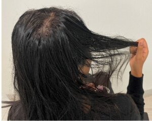
A 25-year-old woman presents to your office noticing increased hair in her shower drain and on her pillow for the past two months. She is concerned that she is losing her hair and continually examines her hair throughout the day.
Question 1: What is important to establish first?
- The duration of hair loss.
- The presence of scalp symptoms.
- Is she reporting hair thinning or shedding?
Answer C. While these are all important questions, first, we should distinguish between hair thinning and hair shedding. Both can present with decreased hair volume. Hair “thinning” refers to the decreasing size of the individual hair shafts. This occurs in androgenic alopecia. It is an androgen-dependent phenomenon where the hair progressively decreases in width and length – a process referred to as miniaturization. The reduced length is due to a shortening of the hair cycle, and thus there is more hair cycling through the telogen phase, where hair falls out, allowing for a new cycle to begin. This might seem like real “shedding,” but in this case the hair is finer (i.e., miniaturized). Interestingly, some real telogen shedding (loss of full-thickness terminal hair) can often be seen in the early stages of both male and female pattern hair loss. This may be due to the low-grade inflammatory process that occurs in these conditions, but the actual etiology is still unknown.
In true hair “shedding” (the medical term is “telogen effluvium”), increased numbers of full-thickness hairs are lost due to a premature shift of hairs from anagen into telogen phase (i.e., a higher number of hairs entering telogen all at once). This can be due to a wide variety of systemic “stressors” that include drugs, trauma, medical conditions, and psychological stress. The hypothesized reason is that the body shifts the growing (anagen) to a resting (telogen) state to conserve energy to deal with stress. The telogen hair will typically begin to fall out approximately 1-3 months later.
To state the difference in a slightly different way, in true “thinning” (caused by androgenetic hair loss), the patient first notices patterned thinning (i.e., thinning in the temples and/or crown) or he/she first notices decreased hair volume. In true shedding, the patient first notices significantly more hair falling out and then a decrease in hair volume. Of course, both androgenetic alopecia and telogen effluvium are extremely common and can co-occur.
As an aside, primary scarring alopecia can cause both thinning and shedding, but they are associated with distinctive changes in the scalp. (See Case #2.)
Question 2: If the patient has increased “thinning,” what are the most important initial questions to ask?
- What is the duration of your hair loss?
- Does anyone in your family have hair loss?
- Do you have any scalp symptoms?
- All the above.
Answer D. All the above. As discussed in question 1, increased thinning is the hallmark of androgenetic alopecia in men and women due to the miniaturization of hair follicles. The duration gives you a sense of the hair loss’s speed and probable future severity. A family history points to a genetic component of the disease. Symptoms of soreness, burning, or itching are more frequent in scarring alopecia than in nonscarring alopecia. However, mild scalp symptoms, such as scalp tenderness or tingling, can also occur in androgenetic hair loss (possibly due to the low-grade inflammation associated with androgenic alopecia. See Case # 13 for more details on androgenic alopecia.)
Question 3: If the patient has increased “shedding,” what are the most important initial questions to ask?
- Have you had any changes in your medications?
- Any recent hospitalizations, especially those that required general anesthesia?
- Any new medical problems?
- Any recent significant emotional stress, i.e., job loss, divorce, or loss of a close family member?
- All the above.
Answer E. These systemic stressors may lead to increased shedding/telogen effluvium. (See Case # 13 for more details on telogen effluvium.)
Question 4: How do you first approach the patient in the physical exam?
- Examine the patient from the front.
- Examine the patient from the back.
- Examine the patient from the top.
Answer A. Although all areas are important to examine, we recommend starting from the front when examining the patient. The frontal view gives you a good look at the patient’s disposition, grooming, and hair loss classification. It also allows you to inspect their facial skin and eyebrows. For instance, temporal facial papules and loss of eyebrow hair may suggest the diagnosis of frontal fibrosing alopecia. Jawline acne and hirsutism may indicate polycystic ovarian syndrome. Also, scarring from traction hair loss, seen with tight hairstyling practices, often presents first at the hairline.
Question 5: When would you perform a hair pull and/or tug test (indicate all that apply)?
- When you are evaluating for active shedding.
- When there is a concern about genetic abnormalities.
- Perform on every patient.
Answer: All the above. The hair pull test is a simple diagnostic test that can help indicate active hair shedding. Genetic abnormalities often manifest as changes in the hair shaft leading to hair fragility, which can be tested with the tug test. The hair pull test and tug test (see below for details) should be performed as part of a routine examination of the hair and scalp.
Question 6: What is true about correctly performing a hair pull test? (indicate all that are true)
- Have the patient vigorously wash and brush 24 hours before the examination.
- Grasp approximately 50-60 hairs between the thumb and index finger. Pull firm and gently away from the scalp.
- Look at whether the hair that comes out is full-thickness (terminal), finer (miniaturized) or broken (hair shaft defect).
- More than 5-6 hairs constitute a positive result.
Answer: All the above. It is important to standardize the hair pull test for reproducible results, which includes ensuring that no shampooing or washing of the hair has occurred for the past 24 hours. Otherwise, the test may be falsely negative. If someone has not shampooed or brushed for more extended periods, the hair pull test can be false positive. This test should always be performed on multiple regions of the scalp (especially the top and back of the scalp or any area where the scalp appears abnormal). Telogen effluvium, anagen effluvium, loose anagen syndrome, early androgenetic alopecia (in both men and women), and active alopecia areata can all result in a positive hair pull. However, the hair that comes out will look different in each case. In telogen effluvium, the end of the hair shaft has a small bulb (making the hair look club-like); in anagen effluvium, there is a gelatinous cylinder (internal root sheath) at the end; and in early AGA, the hair is partially miniaturized. In alopecia areata, the hair pull may be positive in the active border, with diagnoses being made by closer examination of the scalp.
Question 7: How do you perform a hair tug test?
- Same as above. It is another term for the hair pull test.
- Grasp a section of hair and hold it with two hands (one near the root and one near the tip), then tug to see if any strands break in the middle.
Answer B. This test gives the dermatologist information about the brittleness or fragility of your hair strands. This test is useful when one suspects a hair shaft abnormality, which causes hair strands to weaken and break. The extracted hair should then be placed on a white card or sheet of paper to better visualize the hair shafts and determine the percentage of hairs are affected. You can also use a “card test,” where the hair is placed over a white surface while still in the scalp. This will allow you to visualize changes in situ (such as the broomstick appearance of trichorrhexis nodosa).
Question 8: How do you extract hair?
- Extracting hair is when you grab the base of a hair with a clamp or needle holder, quickly yank the hair out, and then place it under significant magnification or a microscope.
- Extracting hair is what you do on a hair pull test.
- Extracting hair is the best way to examine telogen hair.
Answer A.
Question 9: What is the purpose of extracting hair?
- Extracting hair is the only way to see the full length of the hair – whether it is in anagen, telogen or broken from a hair shaft abnormality.
- Extracting hair is the best non-surgical way of examining the root/bulb.
- If you were to suspect a hair shaft abnormality from the hair tug test, then this gives you better clarity by being able to view the entire hair shaft and root under the microscope.
Answer: All the above. Examining extracted hairs under a magnifier or microscope can be helpful in several situations. This includes distinguishing between anagen hairs (found in loose anagen syndrome, anagen effluvium, and primary scarring alopecias) from telogen hairs seen in telogen effluvium. You can also magnify the hair shaft to evaluate for structural hair abnormalities such as trichorrhexis invaginata, monilethrix, trichorrhexis nodosa, etc.
Question 10: What is the main difference between the appearance of anagen and telogen hairs?
- The appearance of the bulb.
- The appearance of the shaft.
Answer A. The best way to distinguish anagen hairs from telogen hairs is by examining the appearance of the bulb. In anagen, the bulb has pigment and a surrounding root sheath. Anagen hairs are seen after a strong hair pull test to pull out normal hair or with a gentle pull in anagen effluvium. In telogen, the bulb has a club appearance and lacks pigment. The lack of pigment is due to the cessation of pigment production at the end of the growth phase. Most fallen hairs are typically telogen hairs. They can also can be obtained using a gentle hair pull test.
Question 11: What should you do next after a hair pull test?
- Use trichoscopy.
- Biopsy the scalp.
- It depends upon the clinical situation.
Answer A. Trichoscopy is a simple, non-invasive technique for visualizing the hair shaft and scalp. Details of the hair shaft, hair follicle opening, perifollicular epidermis, and blood vessels can help point toward different hair conditions. A biopsy should be performed when the diagnosis is in question. Trichoscopic findings can be used to guide the biopsy site. For instance, with lichen planopilaris, find an active location with erythema and perifollicular scale and then incorporate this, as well as some normal appearing skin, in the biopsy.
If you are experiencing gradual hair thinning, please contact our office for an appointment. Call 212-826-2400 or schedule a consult here.


