Cosmetic Surgery Times features Dr. Bernstein’s presentation to the 55th annual meeting of the American Academy of Dermatology in their April 1997 issue.
The article entitled, “Follicular Transplants Mimic Natural Hair Growth Patterns,” describes Dr. Bernstein’s introduction of his new procedure, Follicular Unit Transplantation, to the academy as well as the keys to making the technique successful. Find the complete article below:
Form Follows Function: Follicular Transplants Mimic Natural Hair Growth Patterns
By Neil Osterweil
Contributing Editor
SAN FRANCISCO – In recent years, many hair replacement surgeons have adopted the modem architecture philosophy that “less is more,” moving from the use of hair plugs, to split grafts, to minigrafts and, finally, micrografts. But at least one hair transplant specialist contends that a more appropriate architectural dictum is “form follows function.”
In other words, the surgeon should let the technique fit the head, and not the other way around, suggested Robert M. Bernstein, MD, at the 55th annual meeting of the American Academy of Dermatology.
Dr. Bernstein is an assistant clinical professor of dermatology at the College of Physicians and Surgeons, Columbia University in New York. He described his “follicular transplantation” technique in a meeting presentation and in an interview with COSMETIC SURGERY TIMES.
Natural Hair Groups Used
 “Hair doesn’t grow singly it grows in naturally occurring groups of from one to four hairs. In follicular transplantation, we use these naturally occurring groups as the unit of the transplant,” he told CST.
The typical follicular unit consists of one to four terminal hairs, one or two vellus hairs, sebaceous glands, subcutaneous fat and a band of collagen which circumscribes and defines the unit. In the follicular transplant technique, the follicular unit is carefully dissected and removed, and then the intervening skin is discarded. This enables the donor site to be small, allowing implantation through a small needle poke. Because trauma to the recipient sites is minimal, the entire procedure can be performed at one time. Dr. Bernstein and colleagues have implanted as many as 3,900 follicular units in a single, 1 day session.
Keys to the follicular transplant technique are:
• Identify the patient’s natural hair groupings and isolate the individual follicular units – Hair groupings are assessed with an instrument called a densitometer, and the average size of a person’s groups can be easily calculated. This information is critical in the planning of the transplant. The density of hairs in an individual measured as the number of hairs per square millimeter of skin is quite variable, but the density of follicular units is relatively constant within individual races.
Most people of Caucasian ancestry have a density of approximately one group per millimeter; people of Asian and African descent tend to have slightly less dense growth patterns, although the characteristics of the person’s hair (such as wavy or wiry hair), can give a full appearance even with low density.
If a patient has an average hair density of two, he will receive mostly two hair implants, with some one-hair and three hair implants mixed in. “If you try to make the groups larger than they occur naturally, they will look pluggy. If you try to make them smaller than they naturally occur, they’re not going to grow as well, because each group is actually a little biologic machine that makes the hair — it’s an anatomic unit. If you break it up it just doesn’t grow as well,” Dr. Bernstein observed.
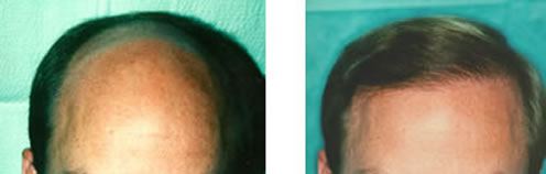
A 38-year old man with a Norwood Class 5A/6 hair loss pattern undergoes a single procedure of 2,500 follicular implants. The result 11 months later. (Photos courtesy of Robert M. Bernstein, MD)
• Harvest meticulously – The acquisition and preparation of grafts must be carefully performed to ensure success for this demanding technique. Highly trained, skilled assistants are essential to the success of the procedure. Dr. Bernstein noted that he uses a highly trained team of up to 10 assistants to produce the implants for a single case. “The assistants, who range from medical technicians to registered nurses, are such an integral part of the procedure that they must become expert in their specific tasks for the surgery to be successful.” The physician must be able to skillfully harvest the donor strip and must be able to make accurate judgments about the size of grafts intra-operatively and adjust the technique accordingly. Dissection and placing of the follicular units is the most labor intensive part of the procedure.
• Design the recipient area well – The recipient sites are carefully distributed so that a natural looking pattern is maintained throughout the recipient area. An important consideration for this stage of the procedure is to “frame the face and spare the crown” so those facial features are kept in correct proportion. A common mistake in hair replacement, said Dr. Bernstein, is to create a hairline that is too high thereby elongating the forehead and accentuating, rather than minimizing, the patient’s baldness. It is also important to avoid or eliminate contrast between the implants and surrounding skin by creating a soft transition zone of single hairs and to have the hair emerge from the scalp at natural angles.
Procedure Lowers Cost
Although the procedure is highly labor intensive, it can actually be less expensive than conventional hair replacement surgery, because it can be performed in a single, but lengthy, session.
“It is also much more efficient and conserves donor hair much better than conventional hair transplants. Every time you make an incision in the person’s scalp you waste some hair and make the remaining hair more difficult to remove. Accessing the donor area just once or twice will increase the total amount of hair that is available for the transplant,” Dr. Bernstein told CST.
“In the very near future, the procedure will be improved and made more affordable with automated instruments that will enable the surgeon to make sites and implant the hair in a single motion. This will also decrease the possibility of injury to the implants by reducing handling and keeping the grafts uniformly cool and moist. It is possible that someday hair follicles may be cloned to provide a virtually unlimited supply of custom follicular units, but until then the finite nature of a person’s donor supply must be respected,” concluded the doctor.
Posted by Robert M. Bernstein M.D.