
Robert M. Bernstein, M.D. and William R. Rassman, M.D.
Hair Transplant Forum Intl. 2012; 22(4): 128-130.
In 1984, Dr. O’Tar Norwood and Dr. Richard Shiell proposed the concept of an X Factor, some unknown factor, or factors, which can lead to sub-optimal growth following a hair transplant. ((Norwood O, Shiell R. X-Factor; Unexpected Unexplained Reduced Hair Growth. In: Hair Transplant Surgery, 2nd ed. O’tar T. Norwood and Richard C. Shiell, eds. Springfield, Illinois: Charles C Thomas, 1984; 134.)) Over the years, as these “unknown” factors have been identified and addressed, graft survival has improved. Some of the factors that have been responsible for the improvements in growth have included; (a) minimizing surgical transection of the follicles at the time of harvesting, (b) preventing graft desiccation, (c) reducing the time grafts are held outside the body, (d) improving holding solutions and, (e) decreasing crush injury during placing. The purpose of this paper is to suggest additional manipulations to increase the survival of grafts in FUE hair transplant procedures.
FACTORS AFFECTING GRAFT SURVIVAL
Grafts In-Vitro
In 1996, Dr. Bobby Limmer underscored these concerns by reporting decreased survival when grafts spend prolonged periods of time outside the body. ((Limmer BL. Micrograft survival. In: Hair Replacement Medical and Surgical. D. Stough and R. Haber, eds. St. Louis: Mosby, 1996; 147-9.)) Following that work, there has been a significant effort to streamline the surgical process, so that grafts could be planted as quickly as possible; a particular concern in large surgical sessions. Graft survival has also been enhanced by using metabolically optimized holding solutions and keeping grafts in a chilled environment while awaiting placement. ((Cooley JE. Ischemia-Reperfusion Injury and Graft Storage Solutions. Hair Transplant Forum Intl. 2004; 14(4): 121, 127, 130.)), ((Parsley WM, Perez-Meza. Review of Factors Affecting the Growth and Survival of Follicular Grafts. J. Cutan Aesthet Surg. 2010; 3(2): 69-75.)), ((Cole JP, Reed WM. The optimal holding solution and temperature for hair follicle grafts. Hair Transplant Forum Intl. 2012; 22(1): 17-21.))
Grafts In-Vivo
One of the first responses to the creation of a recipient wound is the formation of a clot. This has great adaptive value for an organism, where the immediate containment of blood loss takes precedence over the healing process. However, a response that is crucial for the preservation of a species, may present a problem for newly transplanted grafts that have been sitting in a holding solution outside the body.
In the 1995 publication “Follicular Transplantation,” Bernstein and Rassman hypothesized that a “snug fit” of newly placed follicular unit grafts in the recipient wounds would minimize dead space and reduce clot formation. This, in turn, would facilitate the diffusion of oxygen and nutrients into the newly transplanted tissue and promote optimal graft survival. ((Bernstein RM, Rassman WR, Szaniawski W, Halperin A: Follicular Transplantation. Intl J Aesthetic Restorative Surgery 1995; 3: 119-32.)) The crusts that form on the surface of a newly coagulated wound may also serve as a nidus for superficial bacterial colonization and incite an inflammatory reaction required for their removal; therefore, manipulations that minimize them would also be beneficial for the grafts.
It has long been assumed that once grafts are transferred from the cold, hypoxic environment of the holding solution, into the warm milieu of the human scalp, their metabolic needs would immediately be fulfilled. However, the initial environment of the recipient site may not be as immediately hospitable to grafts as previously believed. Although clotting and changes in vascular permeability occur within minutes of wounding, the reabsorption of the clot, the mobilization of an inflammatory infiltrate, angiogenesis, the formation of granulation tissue, and collagen deposition takes hours to days to develop. ((Wasserbauer S. Wound healing for the hair transplant surgeon. Hair Transplant Forum Intl. 2012; 22(2): 37, 42-44.))
During the first 24 hours following recipient wound creation, a flurry of biologic activities take place that facilitates healing. These include the migration of platelets with subsequent release of cytokines, growth factors and pro-inflammatory proteins (histamine, serotonin, kinins, prostaglandins, etc.) that increase blood vessel permeability and stimulate cell migration. Allowing these processes to begin before implantation of the grafts should be beneficial to their healing and subsequent growth. ((Stadelmann WK, Digenis AG, Tobin GR. Physiology and healing dynamics of chronic cutaneous wounds. Am J Surgery, 1998; 176(2): 26-38.)), ((Schremi S, Szeimies R, Prantl L, et. al., Wound healing in the 21st century, J Am Acad Dermatol, 2010; 63: 866-81.)), ((Gantwerker EA, Hom D: Skin: Histology and Physiology of Wound Healing, Facial Plastic Surgery Clinics of N.A. Aug 2011; Vol 19: 3: 441-453.))
Graft Placement
Another aspect of the hair transplant that can adversely affect the survival of grafts is crush injury during placement. When grafts are placed in newly made recipient sites they tend to pop up (or completely out) due to active blood flow and the slippery nature of fresh wound edges. This necessitates re-insertion, subjecting the grafts to additional injury. In addition, the popped grafts sitting above the skin surface are more subject to desiccation and hypoxic injury compared to grafts still in their chilled, holding solution. Over time, however, the bleeding subsides and the wound edges become more “sticky” due to activation of the coagulation cascade. This enables grafts to be placed more easily so that the latter part of the placement process proceeds with greater ease than the initial phases.
Risks to Optimal Growth
In sum, after follicular unit grafts have been isolated in FUT (through strip harvesting and microscopic dissection) or FUE (via direct extraction), there are three additional phases of a hair transplant where grafts are subject to metabolic and physical stresses; in their holding solution, during graft insertion, and after being placed into a fresh recipient wound. In each of these steps, follicular unit grafts are at risk to factors that may contribute to sub-optimal growth.
COMPARING FUT AND FUE PROCEDURES
The typical sequence for a Follicular Unit Transplant performed via strip harvesting (FUT) or direct extraction (FUE) is:
Standard Hair Transplant
Step #1: Obtaining follicular units
Step #2: Site creation
Step #3: Graft placement
In FUT, follicular units are obtained through the stereo-microscopic dissection of a donor strip. ((Limmer BL. Elliptical donor stereoscopically assisted micrografting as an approach to further refinement in hair transplantation. Dermatol Surg 1994; 20:789-793.)) Since this is an in-vitro process, graft dissection can proceed simultaneously with the in-vivo steps of site creation and graft placement. In FUE, grafts are removed directly from the donor area. ((Rassman WR, Bernstein RM, McClellan R, Jones R, et al. Follicular Unit Extraction: Minimally invasive surgery for hair transplantation. Dermatol Surg 2002; 28(8): 720-7.)) Since this is an in-vivo process, it makes it difficult for graft extraction to be accomplished at the same time as the other in-vivo steps of site creation and graft placement. As a result, in standard FUE procedures, there is a delay from the time grafts are extracted from the body until they can be placed into the recipient sites.
In FUT, the microscopic dissection can be done in parallel, with many dissectors working simultaneously to complete the process quickly. In FUE, the logistical constraints of extraction limit it to being performed by one person at a time, significantly increasing the duration of this step of the procedure. ((Harris JA. The SAFE System: New Instrumentation and Methodology to Improve Follicular Unit Extraction (FUE). Hair Transplant Forum Intl. 2004; 14(5): 157, 163-4.))
In FUT, the doctor first decides on a specific number of grafts, harvests a donor strip estimated to contain those grafts and then makes recipient sites based on the number actually obtained from the donor strip. ((Bernstein RM, Rassman WR: Follicular Transplantation: Patient Evaluation and Surgical Planning. Dermatol Surg 1997; 23: 771-84.)) In FUE, the doctor can harvest the exact number of grafts needed from the donor area, so there is usually no need to wait until the extraction is done to determine the number of recipient sites needed.
With some manual FUE techniques, it is possible to do all, or part, of the extraction process with the patient in a sitting position, so that at least part of his recipient scalp is accessible for concomitant site creation and placing. With current robotic technology, however, the movement of the robotic arm and the techniques used to stabilize the head preclude the extraction of follicular units and the creation of recipient sites from being performed at the same time. ((ARTAS™ System manufactured by Restoration Robotics, Inc. (Mountain View, CA).)) (Figure 1.)
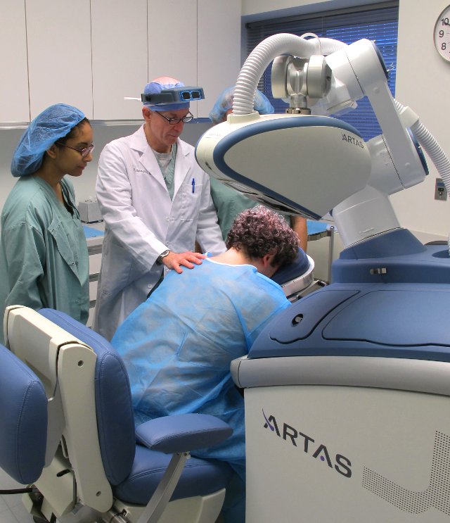
Figure 1. Positioning the patient for robotic FUE
SUGGESTIONS
Changing the Sequence in FUE
The delay in FUE procedures, from the time grafts are extracted from the body until they can be placed into the recipient sites, can be reduced by simply creating the recipient sites prior to extraction. The problem in FUT, of not knowing exactly how many sites are needed, is a non-issue in FUE since the doctor can harvest the exact amount desired from the donor area.
Therefore, FUE procedures lend themselves to easily reversing the normal FUT sequence of graft (strip) harvesting followed by site creation. By making recipient sites first, the time grafts are out of the body will be reduced.
These “pre-made” recipient sites will also exhibit less bleeding than newly created sites and will exhibit the stickiness that makes older sites easier to place grafts into and have less popping. Thus, besides allowing the placing step to proceed more quickly, pre-making sites will reduce the risk of mechanical injury inherent in repositioning elevated grafts.
Another advantage of creating sites before harvesting follicular units is that it provides time for the removal of crusts from the surface of the wound prior to implantation. The removal of these crusts will decrease post-operative inflammation and promote wound healing. A final benefit of reversing the order is that these pre-made recipient sites will start to heal, perhaps making them a more fertile bed for the newly implanted grafts.
The authors suggest that the following basic sequence be used for Follicular Unit Extraction procedures:
FUE with Pre-Made Sites
Step #1: Site creation
Step #2: Obtaining follicular units
Step #3: Graft placement
Delaying Graft Extraction
With large sessions, these authors and others ((Bauman A, Harris J. (Personal communications) )) sometimes extend the FUE procedure over a two day period simply because the more time consuming, large FUE procedures cannot be completed in a single session. Given the described advantages of pre-making recipient sites in an FUE procedure, the question arises if there should be a deliberate delay between site creation and the harvesting steps by creating recipients sites the day prior to harvesting and graft placement, even if the size of the session does not necessitate this.
We have suggested that a recipient wound may become a more hospitable bed for grafts and easier to place over time. This optimal time is, at present, unknown. However, with our knowledge of wound healing and our experience with consecutive day procedures, it seems that making recipient sites the day prior to graft harvesting may offer some advantages over procedure completed in one session.
FUE with Pre-Made Sites and Delay
Step #1: Site creation
Step #2: Deliberate delay (up to 24 hrs)
Step #3: Obtaining follicular units
Step #4: Graft placement
The authors considered the possibility that placing grafts into a recipient site further along in the healing process might increase the risk of re-perfusion injury.4,5 This possibility was deemed unlikely, however, as the second day post-op, the grafts are still relying on diffusion, rather than neo-vascularization, for their oxygenation. Therefore, the factors normally associated with re-perfusion injury would not seem to apply.
The expediency of one-day surgery for the physician and the convenience to the patient having a hair transplant completed in a single session are obvious advantages to one-day procedures. ((Bernstein RM, Rassman WR. Anderson KW. Follicular Unit Extraction Megasessions: Evolution of a technique. Hair Transplant Forum International 2004; 14(3): 97-99.)) However, for larger sessions of FUE, where thousands of grafts have to be negotiated, it may be worthwhile to consider a two-day protocol if it could be shown to facilitate growth.
Conclusion
We have examined two methods of improving the follicular unit extraction procedure. The first decreases the time grafts are held in-vitro by pre-making recipient sites prior to the hair being harvested from the donor area. The second adds additional time between site creation and graft harvesting/placement to allow recipient site healing to progress.
These simple manipulations will decrease the time grafts are out of the body, allow for easier graft placement, allow for crust removal, and possibly create a more fertile bed for the grafts. When performing follicular unit extraction, there is an obvious advantage to making recipient sites before the grafts are harvested. One might also consider delaying extraction to allow the healing process to proceed. The optimal length of this delay, however, remains to be determined.


