
Robert M. Bernstein, M.D. and William R. Rassman, M.D., New York, NY
Hair Transplant Forum International 2001; 11(1): 1, 11-13.
In a collaborative effort in 1998, twenty-one hair restoration surgeons in the ISHRS attempted to “Standardize the Classification and Description of Follicular Unit Transplantation and Mini-Micrografting Techniques.” ((Bernstein RM, Rassman WR, Seager D, Shapiro R, et al.: Standardizing the classification and description of follicular unit transplantation and mini-micrografting techniques. Dermatologic Surgery 24:957-963, 1998.)) It has been the opinion of many physicians who perform Follicular Unit Transplantation exclusively in their practices, that strict definitions of FUT are essential, both to compare different techniques and to enable physicians to clearly communicate with their patients and with each other. However, some feel that strict definitions of Follicular Unit Transplantation are unnecessary, especially with respect to the requirements for single-strip harvesting and stereo-microscopic dissection.
In order to test the validity of the argument that the techniques of single-strip harvesting and stereo-microscopic dissection are essential aspects of Follicular Unit Transplantation, a retrospective study was conducted to examine the effectiveness of obtaining follicular units using different methods of harvesting and dissection in patients undergoing their first hair transplantation procedure.
The Three Techniques Studied
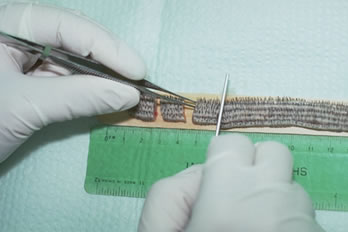
Mini-micrografting Technique
The first technique is the one most similar to that used by mini-micrografters, namely harvesting with a multi-bladed knife (blades set 3mm apart), division into smaller sections (of approximately 1cm), and followed by subsequent dissection with loop magnification (1.5x). (Figure 1) The major difference between this technique and traditional mini-micrografting was that there was a conscious effort to dissect out intact individual follicular units, rather than to merely cut the strips generated by the multi-bladed knife into smaller pieces depending upon the physical size or number of hairs desired (mini-micrografting cut-to size). Hair fragments were not counted in any measurements in the study, but were planted if they were deemed to be of sufficient size to have stability in the recipient site made by an 18g Nokor needle.
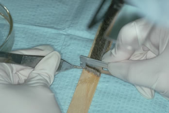
Vertical Sectioning
The second method, that we called “Vertical Sectioning,” was to harvest the donor tissue as one intact strip. This was accomplished by using only the outer blades of the multi-bladed knife. Once the strip was removed, smaller sections were generated by cutting vertical slices at a spacing of approximately 2.5 mm. The pieces generated by this technique were further dissected into individual follicular units under loop magnification. (Figure 2) In this technique as well, hair fragments were often planted, but not counted in the data.

Follicular Unit Transplantation
In the third method, Follicular Unit Transplantation was performed as defined by the 1998 collaborative effort, using single strip harvesting and stereo-microscopic dissection. (Figures 3a, b). Following the removal of the donor tissue as a single strip, the tissue was divided into smaller pieces under stereo-microscopic control through a process called “Slivering.” Individual follicular units were then dissected from these pieces, again using stereo-microscopes.
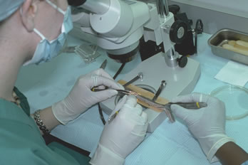

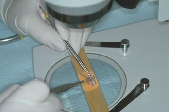

Figure 3b. Subsequent stereo-microscopic dissection into individual follicular units.
Comments on the Techniques
Of importance is that in both the multi-bladed knife technique and the method of Vertical Sectioning, the initial cuts through the strip are performed “blind.” In other words, the surgeon does not have the visual control to avoid splitting follicular units or transecting individual follicles. The theoretical advantage of Vertical Sectioning over that of the multi-bladed knife is that these initial “blind” cuts are vertical rather than horizontal, so that potential damage from not following the incident angle of the emerging hair can be reduced.
The difference between Vertical Sectioning and the “slivering” of Follicular Unit Transplantation is that in the former procedure the sectioning can follow the emerging angle of the hair, but is unable to avoid transecting the randomly placed follicular units, since the cutting of the sections is with a linear motion. This contrasts with the slivering technique of FUT where, under direct stereo-microscopic control, the dissector’s blade passes around follicular units, both to isolate them and preserve their structure.
There are two common ways to perform “slivering.” In the first, slivering is used to divide the donor strip into smaller pieces of approximately 2mm in thickness. Follicular units are then generated from these pieces by further slivering. In the second method, slivering is used to dissect the donor strip directly into actual “slivers” that are each 1-follicular unit (approximately 1mm) wide. The follicular units are then isolated by simply trimming the excess tissue between them. In our study the former technique was used. The important aspect of the procedure is that in both cases, every aspect of the technique is performed under stereo-microscopic control to keep follicular units intact and avoid follicular transection.
It has been recently been pointed out by Stough (personal communication), that some dissection instruments such as the Mantis microscope are not true “stereo-microscopes” and, therefore, the definition should be more inclusive. This is an important point, and any dissecting device that has comparable magnification and illumination to the Meiji Stereo-microscope as originally used by Limmer ((Limmer BL: Elliptical donor stereoscopically assisted micrografting as an approach to further refinement in hair transplantation. Dermatologic Surgery 20:789-793, 1994.)) could serve as a logical substitute. In fact, the methodology used in the year 2000 data in this study included a Mantis microscope for the slivering aspect of the procedure and Meiji microscopes for subsequent dissection.
Stough has also rightly pointed out that, although single-strip harvesting and microscopic dissection are necessary when performing the initial phases of harvesting and dissection when treating Asians with coarse hair, once the slivering process is complete, further dissection can be adequately performed with magnifying loops. For practical purposes, dissection teams facile with the microscope are not at disadvantage to use the more precise dissection throughout the entire procedure; however, this may not be necessary in all cases. The point is that this is another argument against having rigid definitions of FUT methodology requiring “complete” microscopic dissection.
The data in this study was compiled from operative reports completed during the stated periods. The staff’s instructions during this period were to count only largely intact follicles, but to place everything that they deemed to be viable. Grafts that consisted of only transected hairs and/or hair fragments were not counted. Diagrams of different follicular components and follicular unit/split hair combinations were discussed and posted in order to standardize policy and this policy remained relatively unchanged during the study period. Graft sizes were determined by the method of magnification used for the dissection (i.e. loops or microscope) and were recorded by the staff as they came off the dissecting block. It is important to point out, that since this was a retrospective study that spanned 5 years, is was impossible for the criteria determining exactly what was a hair fragment and what was an intact follicle to be uniformly consistent. The data should be viewed in this context. The criteria also differed from our Bilaterally Controlled Study of 19981 where all hair fragments that were deemed to be viable were counted.
The Findings
The results of the study are summarized in the following three tables:

Please note: To exclude atypical cases, the smallest 2.5% and largest 2.5% were not included in the data.
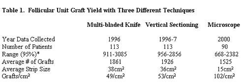
Table 1 shows that the average number of grafts per case decreased from 1,861 grafts per case in 1996 and 1,926 in 1996-7 to 1,525 grafts per case in the year 2000. This occurred in spite of the fact that our goals for the extent of planned coverage remained the same. ((Bernstein RM, Rassman WR, Szaniawski W, Halperin A: Follicular Transplantation. International Journal of Aesthetic and Restorative Surgery 1995; 3: 119-132.)), ((Bernstein RM, Rassman WR: Follicular Transplantation: Patient Evaluation and Surgical Planning. Dermatologic Surgery 23:771-784, 1997.)), ((Bernstein RM, Rassman WR: The Aesthetics of Follicular Transplantation. Dermatologic Surgery 23:785-799, 1997.)) It was also our subjective experience that the clinical results appeared to improve over this period, as measured by the degree of fullness that was achieved from the transplant.
During this same period, the size of the strip that was harvested decreased even more dramatically than the number of grafts we obtained from them, with 37cm2 the average strip size in the 1996 and 1996-7 period and only 15cm2 in the year 2000. Again, this occurred over a period when the clinical results continued to improve, although by the nature of this being a longitudinal retrospective study other aspects of the surgery changed as well.
When one looks at the number of grafts harvested per cm2 one observes that the number of intact follicular units harvested per cm increased dramatically from 49/cm2 in 1996 to 53/cm2 in 1996-7 and to 102/cm2 in the year 2000. Interestingly, the number of follicular unit grafts that we now achieve using stereomicroscopic dissection is almost exactly the number that we have observed with pre-operative densitometry and that would be predicted by the follicular unit constant of 1 follicular unit per cm2.
It is important to note that grafts that consisted of only transected hairs and/or hair fragments were not counted in the study, but were often planted. This will tend to exaggerate the differences in the data between the techniques. The point of the study, however, was to examine the ability of the techniques to dissect follicular units, and not specifically to compare clinical results. When mini-micrografting is performed in clinical practice, essentially all of the harvested tissue is planted, so that most, if not all, transected follicles and hair fragments are transplanted as well. Since a portion of these follicles would be expected to be viable, this would significantly affect the clinical results.


Table 2 examines the actual complexion of the individual follicular units generated by each technique. Using a multi-bladed knife followed by loop dissection, each follicular unit contained an average of 1.74 hairs. When vertical sectioning was used in the initial sectioning of the harvested strip, there were 2 hairs per follicular unit, an almost 15% increase over the multi-bladed technique. With single strip harvesting and complete stereo-microscopic dissection, 2.26 hairs per follicular unit were obtained, 15% more than with vertical sectioning and almost 30% more than when a multi-bladed knife was used. It is interesting to note that the 2.26 hairs per follicular unit obtained with single strip harvesting and stereo-microscopic dissection is similar to that which has been observed using densitometry, indicating that this technique appears to be able to preserve the entire follicular unit.
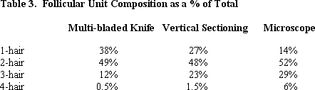

Table 3 examines the type of follicular units generated as a percent of the total yield. It is striking that with the stereo-microscope, 35% of the grafts contained either 3 or 4 hairs, whereas with the multi-bladed knife, only 12.5% contained that many hairs. Even more dramatically, the number of 4-hair grafts obtained with the stereo-microscope was 10 times as many as that obtained with the multi-bladed knife. This helps to explain the greater fullness seen when FUT is performed with the latter technique.
In Perspective
This retrospective study complements a previously published bilaterally controlled study by these authors showing the advantage of microscopic over loop dissection used once the donor tissue has been divided into strips with a multi-bladed knife (as in method 1in this study) or in vertical sections (as in method 2). In the previous study, microscopic dissection produced a 17% greater yield of hair as compared to magnifying loops with transillumination, when the microscope was used for only one aspect of the procedure. ((Bernstein RM, Rassman WR: Dissecting microscope vs. magnifying loops with transillumination in the preparation of follicular unit grafts: A bilateral controlled study. Dermatologic Surgery 24: 875-880, 1998.))
In the conclusion section of that study we wrote “The results of this study show an increase in the yield of follicular unit grafts, as well as the total amount of hair harvested from the donor strip, when using the dissecting microscope as compared to magnifying loops with transillumination. This increase was observed when only the latter part of the dissecting procedure was studied. When complete microscopic dissection is used, the advantage should be even more significant.” The importance of the present study is that it shows what that “significant advantage” is.
It is important to stress that this present study looked at the ability of mini-micrografting techniques to generate follicular units, not to perform mini-micrografting per se. In clinical practice, physicians who perform mini-micrografting do not try to isolate follicular units and, therefore, transplant essentially all of the harvested tissue, so that transected hairs and hair fragments are transplanted as well.
In Follicular Unit Transplantation, transected hairs are also transplanted; however, the issue is moot since, with good technique, few are actually generated. The problem arises when FUT is attempted with techniques that can neither keep follicular units intact nor avoid follicular transection. In this case, attempting to isolate follicular units generates too much hair wastage and negates many of the advantages of the FUT procedure.
When Follicular Unit Transplantation is performed with the proper methodology, i.e. with single- strip harvesting and microscopic dissection, the follicular unit yield is what would be expected if all of the follicular units in the donor tissue were captured. In addition, this technique produces follicular units with a composition comparable to that of normal donor scalp.
Conclusion
This study offers strong support for the argument that if one wants to perform Follicular Unit Transplantation i.e. “A method of hair restoration surgery where hair is transplanted exclusively in its naturally occurring, individual follicular units” then, for the vast majority of patients, “single strip harvesting and microscopic dissection should be required.”


